Pulmonary angiography is an X-ray of the blood vessels that supply the lungs. It is used to find a blood clot, also called a pulmonary embolism, in these blood vessels. The procedure is often done by a specially trained healthcare provider called an interventional radiologist. Or it may be done by a heart specialist (cardiologist). This type of test is rarely done today. Instead, a CT angiography (CTA) of the chest is typically done to find out if you have a pulmonary embolism.
What to tell your healthcare provider
Before the procedure, be sure to tell your healthcare provider if you:
-
Have any allergies to food, medicines, or contrast dyes (mediums)
-
Have had any recent illnesses and all health conditions you have
-
Are pregnant or think you may be pregnant
-
Are breastfeeding
-
Have an irregular heart rhythm
-
Take medicines, vitamins, herbs, or supplements, including over-the-counter medicines and illegal drugs
Getting ready for your procedure
-
Follow any directions you’re given for not eating or drinking before the procedure.
-
Change into a hospital gown. Remove hair clips, jewelry, dentures, and other metal items that could show up on the X-ray.
-
Go to the bathroom to empty your bladder just before the procedure begins.
-
Plan to have a friend or relative available to drive you home.
During your procedure
-
Lie down on the X-ray table. An IV (intravenous) line is put into a vein in your hand or arm. You will be given fluids or medicines through the IV.
-
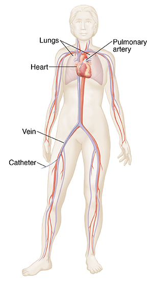
Medicine is put into the skin over your groin or arm to numb it. A needle is then put into a blood vessel near the groin or your arm. The needle is then replaced with a thin, flexible tube called a catheter.
-
Using X-ray images (fluoroscopy) as a guide, the healthcare provider moves the catheter through blood vessels and your heart to the pulmonary artery. This is the artery that carries blood from the right side of the heart to your lungs.
-
X-ray dye, also called contrast medium, is injected into your artery through the catheter. This dye helps the blood flow in your lungs show up better on X-rays. You may feel warmth when the dye is injected.
-
X-ray images are then taken. Stay as still as you can while the X-rays are taken. You may be asked to hold your breath for 10 to 25 seconds at a time. The healthcare provider will tell you when to hold your breath and when to breathe.
-
After the X-rays are taken, the catheter is removed. Pressure will be put on the insertion site for 10 to 20 minutes to stop bleeding.
-
The whole procedure may take about 1 hour.
After your procedure
After the procedure, you may stay in the hospital for a few hours or overnight. When you go home:
-
Care for the insertion site as directed.
-
If the groin was used, keep the leg on that side straight for 6 hours after the procedure.
-
Drink plenty of fluids to help flush the X-ray dye out of your body.
Possible risks and complications
-
Infection or bruising around the catheter insertion site
-
Problems because of X-ray dye, including allergic reaction or kidney damage
-
Damage to a blood vessel by the catheter or blood clots forming in the vein that was first entered
-
Pulmonary embolism because blood clots were released from blood vessel walls that the catheter passed through
-
Short-term abnormal heartbeats