Dismiss Modal

Treatment

Hip Arthroscopy: Repairing Synovitis and Arthritis

Choose a preferred language

Synovitis of the hip causes the lining of the joint (synovium) to become inflamed, painful, and swollen. With arthritis, a large amount of the firm, smooth tissue covering the “ball” and “socket” of the hip (cartilage) is damaged. Using only small incisions and special tools, arthroscopy can help fix synovitis and arthritis.

Cross section of hip joint with femoral head moved out of socket. Cartilage lining socket is damaged. Synovium lining joint is inflamed.


In the operating room

Just before surgery, you may be asked several times which hip is to be treated. This is a standard safety measure. In the operating room, you will likely receive medicine (anesthesia) to make you sleep or numb the area.


During the procedure

After you receive anesthesia, your leg is gently pulled to widen the hip joint. Next, the surgeon makes a few small incisions called portals. Through these portals, the surgeon inserts surgical tools, including the arthroscope. The arthroscope sends images of the joint to a screen. These images allow the surgeon to look inside the joint. The joint is filled with sterile fluid to help the surgeon see more clearly.


Repairing synovitis and arthritis

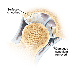
For synovitis, part of the damaged synovium is removed. This may be done by shaving tissue using special tools. For arthritis, the loose or torn cartilage is removed. Or it may be smoothed with a shaver. Arthritis can lead to bone spurs. If bone spurs are present, they may be removed. Once the surgeon finishes, the portals are closed and bandaged. Then you are taken to the recovery room.
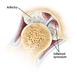
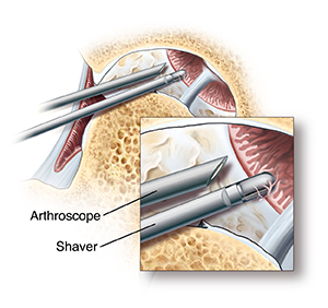
Cross section of hip joint with femoral head moved out of socket. Cartilage lining socket is damaged. Synovium lining joint is inflamed. Arthroscopic instruments are in joint near top of socket. Closeup of arthroscope tip inside joint with shaver removing damaged synovium.Cross section of hip joint with femoral head moved out of socket. Cartilage lining socket is smoothed. Damaged synovium is removed.

© 2000-2025 The StayWell Company, LLC. All rights reserved. This information is not intended as a substitute for professional medical care. Always follow your healthcare professional's instructions.
Related Articles
Read article
Orthopedics
Treating Ganglia

A ganglion is a cystic swelling that is found in overlying joint and tendon sheaths, especially of the wrist. The foot and other extremity areas are sometimes involved. Ganglia can be treated surgically and nonsurgically.

Read article
Orthopedics
Microdiskectomy

During a microdiscectomy, some disk is removed. In most cases, bone must first be removed to expose the damaged disk. The part of the disk outer wall and soft center that presses on the nerve can then be removed. There is usually enough disk remaining to cushion the vertebrae.

Read article
Orthopedics
Ankle Arthroscopy: Conditions Treated

Arthroscopy is used to find and treat ankle problems. These include loose bodies, bone spurs, osteochondritis dissecans (OCD), and synovitis.

Read article
Orthopedics
Treating Bunions

Bunions don't go away. Wearing shoes that fit correctly will often relieve the pain. Bunions that remain painful may need surgery.

Our orthopedic specialists will get you moving pain-free again.

From orthopedic surgeons to physical therapists, our orthopedic team has been built to provide seamless care and support throughout your whole journey.

Browse our specialists