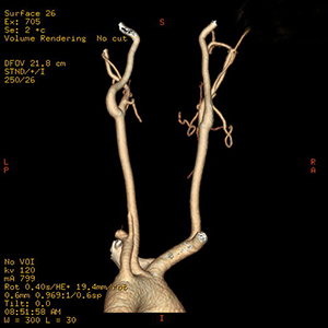
Computed tomography angiography (CTA) is an imaging test. It uses X-rays and computer technology to make detailed pictures of your arteries (blood vessels). Before the test, an X-ray dye (contrast medium) is injected into your vein. The dye makes it easier to see your blood vessels on the X-ray. Pictures are then taken with the CT scanner. A computer turns the images into 2-D and 3-D pictures.
Why CTA is done
CTA may be used to:
-
Check arteries in your belly, neck, lungs, pelvis, kidneys, or brain.
-
Look for a ballooning of the blood vessel wall (aneurysm) or a tear (dissection).
-
Check if a tube (stent) used to keep an artery open is working well.
-
Find damage to your arteries due to injuries.
-
Collect details on blood vessels that supply blood to tumors.
Getting ready for your test
Tell your doctor if you:
-
Have diabetes.
-
Have kidney disease.
-
Are allergic to X-ray dye or other medicines.
-
Are pregnant, think you may be pregnant, or are breastfeeding.
-
Are taking any medicines, herbs, or supplements, including prescription medicines, illegal drugs, and over-the-counter medicines, such as aspirin or ibuprofen.
Follow any directions you are given for not eating or drinking before the CTA. Follow any other instructions from your doctor.
During your test
-
You will be asked to remove any hair clips, jewelry, false teeth, or other metal items that could show up on the X-ray.
-
You will lie down on the scanning table. An I.V. line will be put in a vein in your arm or hand.
-
The scanning table will be correctly placed. The part of your body being checked will be inside the doughnut-shaped CT scanner.
-
One image may be taken first to be sure you are in the correct position for the test.
-
The I.V. will be hooked up to an automatic injection machine. This controls how often and how fast the X-ray dye is injected. The injection may continue during part of the exam.
-
The dye will be put into your vein through the I.V. line. You may feel warmth through your body when the dye is injected.
-
You can’t move while the X-rays are being taken. Pillows and foam pads may be used to help you stay still. You will be told to hold your breath for 10 to 25 seconds at a time.
-
A single scan may take several minutes. You may need more than one scan.
After your test
-
Drink plenty of fluids to help flush the X-ray dye from your body.
-
You may eat as soon as you want to.
Possible risks
All procedures have some risks. A CTA has some possible risks. These include:
-
Problems due to the X-ray dye, such as an allergic reaction or kidney damage.
-
Skin damage from leaking X-ray dye near where the I.V. was put in.


